
Digital X-Rays in Yaletown for Dental Diagnosis
Get digital X-rays in Yaletown for cavities, root issues, and full-mouth imaging. Safe, low-radiation dental diagnostics.
 Rated as “Excellent” (130 reviews)
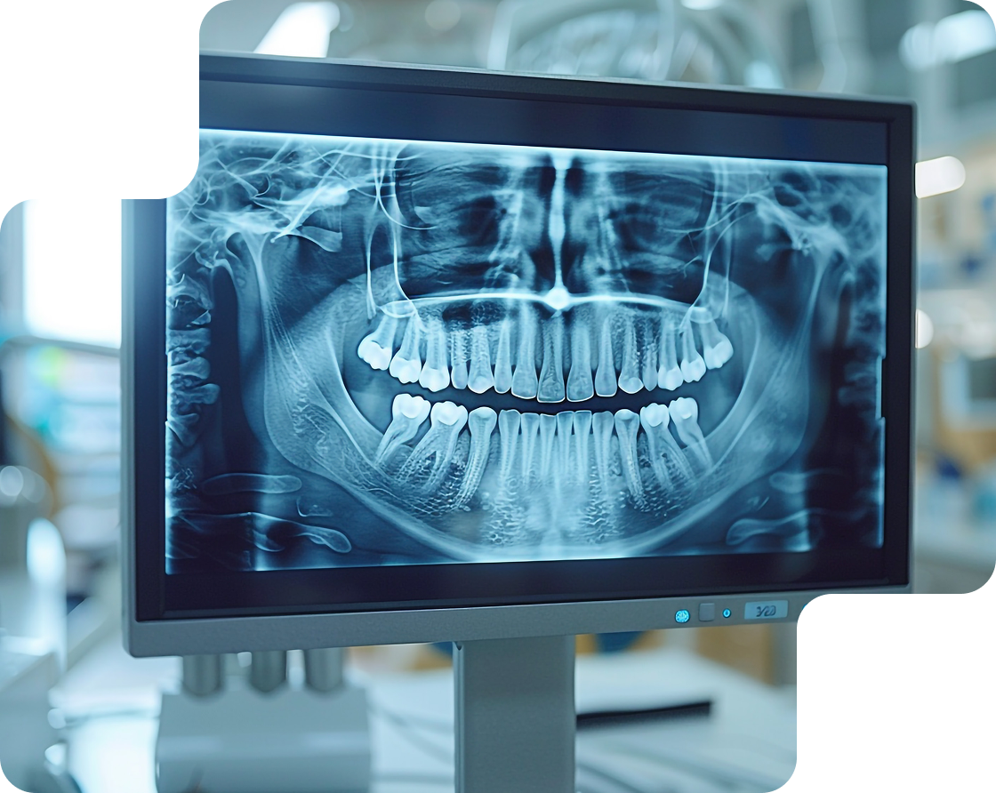
Digital Dental X-Rays in Yaletown — Advanced Imaging for Informed Assessment
Have you ever wondered how a dentist can spot a cavity before you feel it, or how we can diagnose an issue inside a tooth or below the gums without doing anything invasive? That’s exactly what digital dental X-rays make possible. Modern digital imaging gives us clear, high-resolution images in seconds while using dramatically less radiation than old film X-rays.
At Yaletown Dental Boutique, we use three main types of digital X-rays, bitewing, periapical, and panoramic, each designed to reveal different details of your teeth, roots, and jaw.
These images help us detect problems like hidden cavities, early bone loss, root infections, impacted wisdom teeth, and jaw concerns long before they become painful or expensive to fix. The result is faster answers, more accurate treatment planning, and better long-term oral health for you.
Get clarity, catch problems early, and protect your smile. Book your digital X-ray and exam with us now.
Bitewing X-Rays — Spotting Cavities Between Teeth
Bitewing X-rays are the most common type of dental radiograph and are typically taken during routine checkups. A small digital sensor is placed between your upper and lower back teeth, and you bite down gently to keep it in position, this is where the name “bitewing” comes from.
These images capture the crowns of the premolars and molars, along with the spaces where the teeth touch, which are the areas most prone to hidden decay. Because cavities often start between teeth, where a visual exam can’t detect them, bitewing X-rays are X-rays are widely used for early cavity detection and monitoring bone levels around the teeth.
Red, White, or Mixed Patches
Bitewings provide a precise, distortion-free look at the areas most vulnerable to decay and gum disease.
They help dentists identify:
- Cavities forming between teeth (interproximal decay)
- Early enamel demineralization that can still be reversed
- Tartar buildup below the gumline
- Bone loss caused by periodontal (gum) disease
- Gaps, leakage, or breakdown around old fillings and crowns
- In children, development of permanent teeth and spacing
This early insight allows dentists to detect problems before they cause pain, infection, or expensive treatment.
What They Reveal?
Bitewing imaging allows dentists to detect issues long before they become visible or painful.
They can reveal:
- Early cavities that have not broken through enamel
- Weak spots in enamel that can still be remineralized instead of drilled
- Bone changes that signal early gum disease
- Hidden decay underneath old fillings or crowns
- Tooth structural changes that can’t be seen visually
This kind of preventive detection is what helps avoid root canals, extractions, and major restorations later.
When Bitewings Are Taken?
Bitewing X-rays are commonly included in routine dental exams, but how often they’re needed depends on your individual risk for tooth decay and gum disease.
They’re typically taken:
- During standard checkups to screen for cavities and monitor bone levels
- More frequently if you have higher risk factors, such as:
- A history of frequent cavities
- Multiple fillings or crowns
- High sugar intake or frequent snacking
- Orthodontic appliances
- Reduced saliva flow (dry mouth)
For patients with healthy gums and low decay risk, bitewings may be taken less often—every 12 to 24 months instead of annually. Your dentist will tailor the frequency to your needs, following the principle of taking X-rays only when they’re clinically necessary.
Frequency Recommendation
How often bitewing X-rays are needed varies from patient to patient. Dentists do not follow a fixed schedule for everyone, instead, they make the decision based on age, cavity risk, and the current condition of the teeth and gums.
This follows the ALARA principle (As Low As Reasonably Achievable), meaning X-rays are taken only when they are clinically necessary and the diagnostic value outweighs the exposure.
For children and teens, bitewings are often recommended every 6 to 12 months because their teeth are still developing and cavities can progress quickly. Adults who have a history of decay, multiple fillings, frequent snacking, or other high-risk factors usually follow the same 6 to 12 month interval.
Meanwhile, adults with healthy gums, low cavity activity, and good oral hygiene may only need bitewing X-rays every 12 to 18 months.
In every case, the goal is to monitor dental health efficiently while keeping radiation exposure as low as possible. X-rays are taken only when the diagnostic benefit outweighs the risk.

Confident Smiles Start Here — Book Your Visit Today
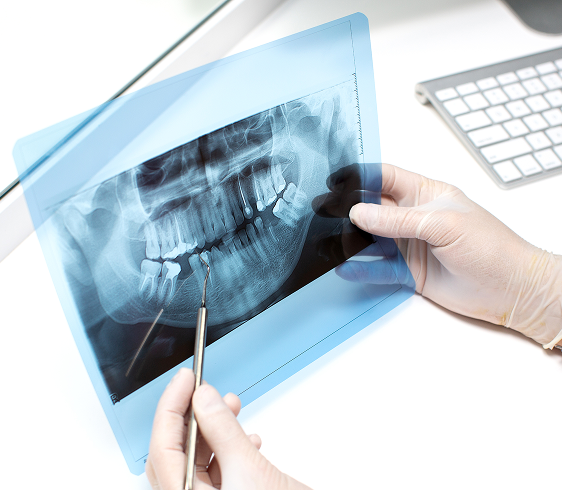
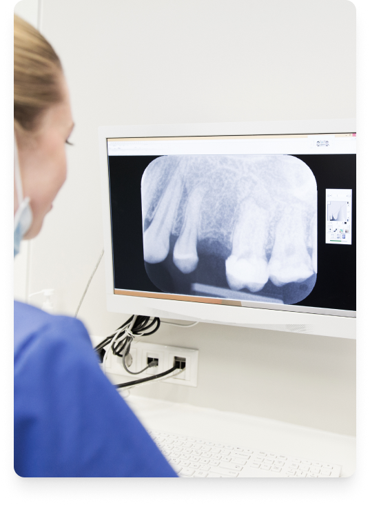
Periapical X-Rays — Examining Roots & Supporting Bone
Periapical X-rays provide a close-up, high-detail view of a single tooth or a small group of teeth, showing everything from the crown down to the root tip and the surrounding bone. Unlike bitewings, which capture only the top portion of the back teeth, periapical images are designed to evaluate what is happening below the gumline, including the root, nerve area, and supporting bone.
These X-rays are essential when there is pain, swelling, or suspected infection in a specific tooth, and they also play a key role in monitoring root canals, implants, and other treatments involving the tooth’s internal structure.
What Periapical X-Rays Show?
Periapicals allow dentists to see the full anatomy of a tooth and the tissue around it, making them the most reliable tool for diagnosing problems at the root level.
They can reveal:
- Abscesses, cysts, or infection at the root tip
- Bone loss associated with gum disease
- Cavities that extend deep toward the nerve
- Cracks or fractures not visible on the surface
- Changes around dental implants or root canal-treated teeth
Because dense structures like enamel and bone appear white while infection or decay appears darker, these X-rays make it easier to distinguish healthy tissue from diseased areas.
Use Cases
Periapical X-rays are ordered when a targeted, tooth-specific diagnosis is needed.
Common reasons include:
- Sudden tooth pain or swelling
- Evaluating a tooth before or after a root canal
- Assessing bone support around a loose tooth
- Checking implant health or healing
- Investigating trauma or a cracked tooth
- Diagnosing deep decay close to the nerve
They are also used as part of treatment follow-up to confirm healing and long-term stability.
How Periapicals Complement Bitewings?
Bitewings detect decay between teeth and monitor bone levels in the back of the mouth, but they do not show the root or deep structures.
Periapicals fill that gap by providing:
- Full root visibility (apex to crown)
- Clear imaging of supporting bone and periodontal space
- Detection of problems that bitewings cannot capture (abscess, root fractures, implant issues)
In practice:
Bitewing : decay detection
Periapical : root, bone, and infection detection
Most full diagnostic exams use both when necessary.
Signs We Look For
On a periapical image, dentists carefully examine:
- The shape and clarity of the root tip
- Dark radiolucent areas indicating infection or cysts
- Bone level and density around the tooth
- Widened periodontal ligament space (sign of trauma or inflammation)
- Irregularities in root canal fillings or implant threads
- Presence of calculus below the gums
Anything that appears darker or irregular around the root usually signals an area of concern.
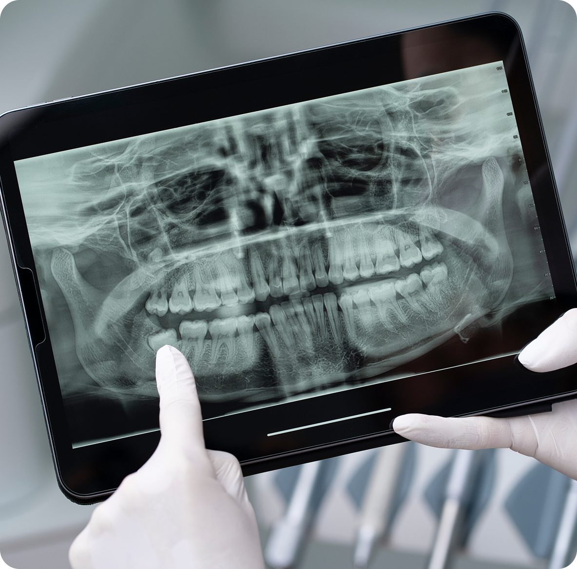
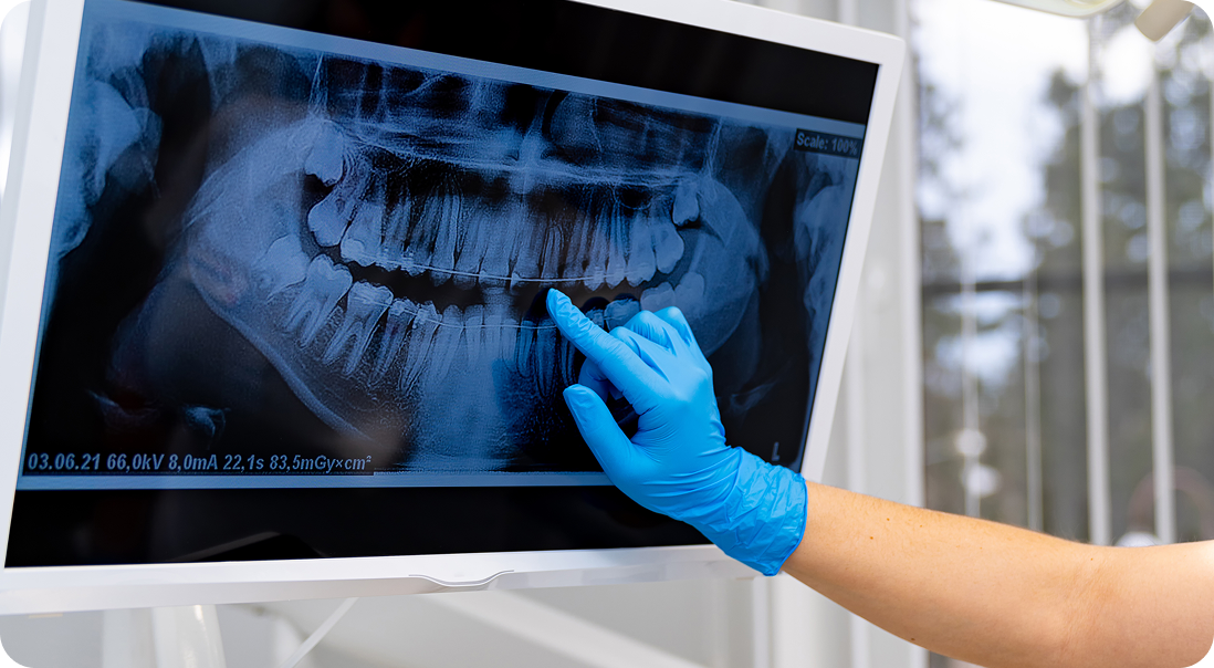
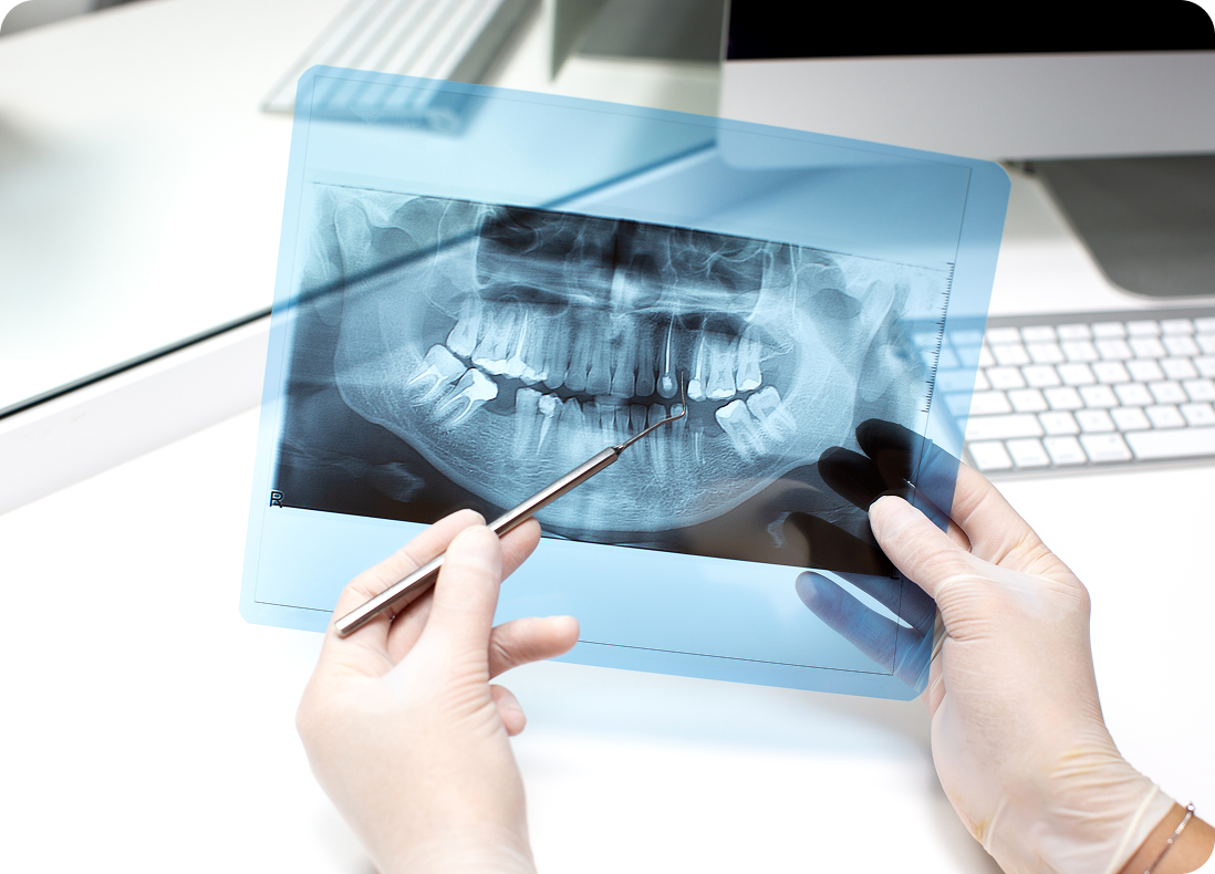
Panoramic X-Rays — Full-Arch Overview in One Shot
A panoramic X-ray is a two-dimensional digital image that captures your entire mouth in a single scan, including all teeth, upper and lower jaws, TMJ joints, sinuses, and surrounding bone structures.
Unlike bitewing or periapical X-rays, the sensor is not placed inside your mouth. Instead, the machine rotates around your head, creating a flat, wide-angle view of your jaw and facial anatomy. Because it shows areas that intraoral X-rays cannot reach, a panoramic scan is often used when a bigger, structural overview is needed instead of a tooth-by-tooth close-up.
Documentation and Photographic Monitoring
Panoramic X-rays are commonly used by dentists and oral surgeons because they cover a much larger area than bitewing or periapical images.
They are typically taken to:
- Plan dentures, braces, extractions, or dental implants
- Evaluate the position of wisdom teeth and other impacted teeth
- Monitor TMJ joint changes or jaw disorders
- Check the progression of gum disease or bone loss
- Diagnose cysts, fractures, or irregularities in the jaw
- Assess patients who are gag-sensitive and cannot tolerate intraoral sensors
Because they show both jawbones at once, panoramic X-rays are also useful when a dentist needs a structural overview instead of a tooth-level close-up. They also support full-jaw screenings and oral pathology checks at our Yaletown clinic.
with Dr. Hadianfar
Dr. Sasan Hadianfar, General Dentist

Safety, Radiation & Why Digital Is Better
Concerns about radiation exposure are common, and understandably so. At Yaletown Dental Boutique, digital dental X‑rays are used because they provide essential diagnostic information while keeping radiation exposure as low as reasonably achievable.
Modern digital imaging technology allows dentists to detect cavities, bone loss, infections, and structural changes early, often before symptoms appear, using significantly lower radiation than traditional film X‑rays. This makes digital radiography a safe and effective diagnostic tool for both adults and children when used appropriately.
Typical Radiation Levels Compared to Film
Digital dental X‑rays expose patients to substantially less radiation than older film-based systems. In many cases, digital sensors reduce radiation exposure by up to 70–80% compared to conventional film X‑rays, while producing clearer and more detailed images.
To put this into perspective, the radiation from common dental X‑rays is very low and comparable to everyday background exposure. For example, a standard set of bitewing digital X‑rays delivers a dose similar to what a person receives from natural environmental radiation over a short period of daily life.
Because digital images are captured instantly and with greater sensitivity, fewer retakes are required, further minimizing exposure while improving diagnostic accuracy.
Protective Measures in Our Clinic (Lead Apron, Thyroid Collar)
Radiation safety at Yaletown Dental Boutique is built into every step of the imaging process. When X‑rays are taken, protective measures are used to limit exposure to non-target areas. These include:
- Lead aprons to shield the body from scatter radiatio
- Thyroid collars when clinically appropriate, especially for children and young adults
- Precise positioning and trained technique to avoid unnecessary retakes
In addition, X-rays are never taken routinely without reason. Each image is prescribed based on clinical need, age, risk factors, and current oral health status. This helps ensure patients receive only the imaging necessary to support clinical assessment and care planning.
Regulatory Guidelines (Health Canada, CDA Standards)
All dental radiography protocols at Yaletown Dental Boutique follow national safety guidelines established by Health Canada and the Canadian Dental Association.
These guidelines emphasize:
- The ALARA principle (As Low As Reasonably Achievable)
- Justified use of radiographs based on individual clinical need
- Proper equipment maintenance and staff training
- Patient protection through shielding and exposure control
By adhering to these standards, digital X‑rays remain a safe, regulated, and essential part of modern preventive and diagnostic dental care.
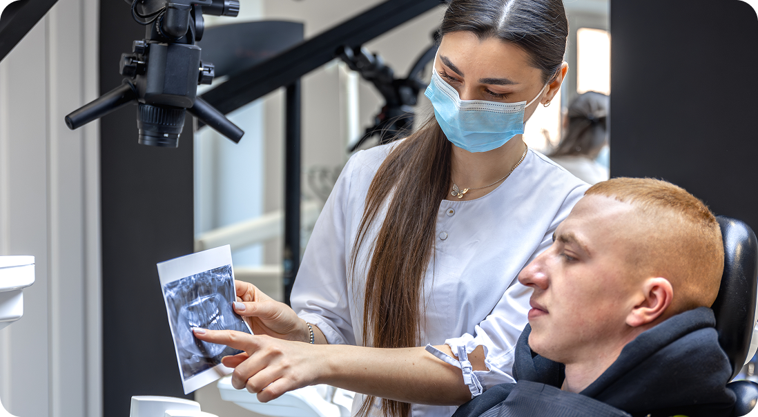
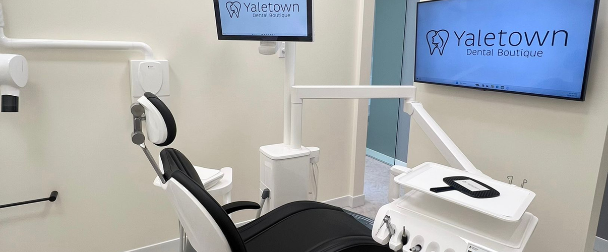
How Digital X-Rays Integrate into Dental Care at Yaletown?
Digital X-rays are not used in isolation, they are fully integrated into how dental care is delivered at our Yaletown clinic. From diagnosis to treatment planning, digital imaging supports clearer communication, more informed decisions, and a smoother overall patient experience.
In addition to standard 2D digital X-rays, our clinic also offers CBCT (Cone Beam Computed Tomography) for advanced 3D imaging when detailed views of bone, roots, or complex anatomy are needed.
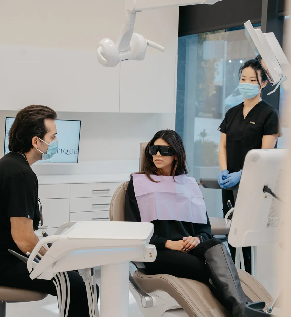
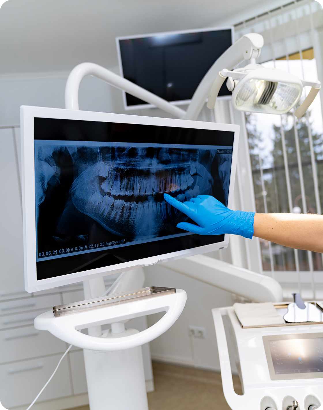
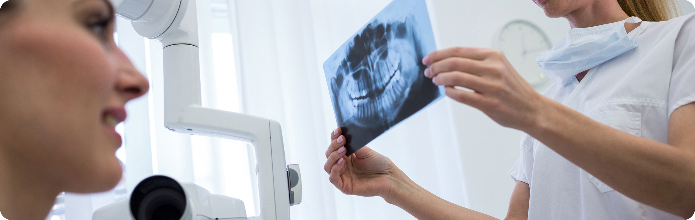
Immediate Image Access for Patient Education
Digital X-ray images appear on screen within seconds of being taken. This allows your dentist to review the images with you in real time, point out specific areas of concern, and explain what’s happening in a clear, visual way.
Seeing the images yourself often makes diagnoses and treatment recommendations easier to understand, helping you make informed decisions about your care.
Choosing the Right X-Ray Type for Your Needs
Not every dental concern requires the same type of imaging. The goal is to select the appropriate X-ray, whether tooth-level detail or a full-arch overview, to support clinical assessment while keeping radiation exposure to what is clinically necessary.
CDCP Patients
Treatment under the Canadian Dental Care Plan is not fully covered. A co-payment will apply.

Why Get Your Digital X-Rays at Yaletown Dental Boutique?
Choosing where you have your dental X-rays taken matters. The quality of the imaging, the experience of the team, and how those images are used all play a role in supporting clinical assessment and treatment planning.
Book Your Digital Dental Imaging in Yaletown
Booking digital dental X-rays at our Yaletown clinic is simple and flexible. Whether you’re visiting for a routine check-up or need imaging for a specific concern, our team makes scheduling and coordination easy.Whenever your plan allows it, we direct bill insurance providers to minimize out-of-pocket costs. Some plans do not permit direct billing, in which case our team will review coverage details with you in advance and provide clear cost estimates before any X-rays are taken.
Business Hours
- Tuesday – Friday: 9:00 am – 5:00 pm
- Saturday: 9:00 am – 4:00 pm
- Closed Sunday & Monday
FAQs
What are digital dental X-rays?
Are digital X-rays safe?
Do I need X-rays at every dental visit?
How long does a digital X-ray appointment take?
Will I feel anything during the X-ray?
Can digital X-rays detect problems early?

Request an Appointment Now!