
At a Glance:
CBCT scans give your dentist a 3D view of your teeth, jaws, and surrounding structures, something traditional X-rays can’t provide. They’re used when precise detail is needed for implants, wisdom teeth, orthodontics, or complex root canals. The scan is quick, painless, and safe, with radiation levels far below those of a medical CT.
In dentistry, the difference between a good result and a great one often comes down to what your dentist can see before treatment begins. Standard X-rays show shadows and outlines; CBCT scans reveal a full 3D landscape of your teeth, jaw, and surrounding structures.
That clarity is why your dentist may recommend this advanced imaging. It’s not about extra tests or extra cost; it’s about precision, safety, and planning every step of your care with confidence.
What is a CBCT scan in dentistry?
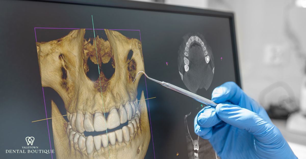
A CBCT scan, short for Cone Beam Computed Tomography, creates a three-dimensional view of your teeth, jaws, and surrounding structures. Unlike standard dental X-rays that flatten everything into a 2D image, CBCT captures depth and detail, giving your dentist a complete picture of what’s happening beneath the surface.
The 3D Imaging Process and Rotating Arm
During the scan, a rotating arm circles your head and collects hundreds of images in about 30 seconds. These images are combined into a detailed 3D reconstruction of your dental anatomy, allowing precise measurements and visualization of even the smallest structures.
Field of View (Single Arch vs. Full Jaw)
Depending on your treatment needs, the scan can focus narrowly on one section, like a single arch, or expand to capture the full jaw and sinus regions. This flexibility makes CBCT valuable for everything from root canal planning to implant placement.
This flexibility allows dentists to capture only the necessary anatomy, a selective approach where the goal is always to use technology responsibly and only when it adds true clinical value.
What conditions can a CBCT scan diagnose?
A CBCT scan isn’t for every patient; it’s used when more detail is needed. By showing precise views of bone, teeth, and tissues, it’s especially valuable in implant dentistry for accurate diagnosis and safe treatment planning.
Here are the main conditions where CBCT provides clear diagnostic advantages:
| When a CBCT Scan Makes a Difference | ||
| Condition | What CBCT Shows | How It Helps the Procedure |
| Dental Implant Planning | Bone Height, Width, Density; Nerve Pathways; Sinus Boundaries | Determines Exact Implant Position and Angulation, Reducing Surgical Risk |
| Impacted or Wisdom Teeth | Root Shape, Tilt, Proximity to Nerve Canal | Helps Plan Safer Extractions and Avoid Nerve Injury |
| TMJ Disorders | Joint Anatomy, Bone Wear, Condyle Position | Assists in Diagnosing Joint Degeneration and Planning Conservative Therapy |
| Root Fractures & Canal Anatomy | Micro-Fractures, Complex Canals, Missed Canals | Improves Accuracy in Root Canal Treatment and Retreatment |
| Cysts, Tumours, Lesions | Size, Shape, Exact Location of Abnormalities | Supports Early Diagnosis and Timely Referral When Needed |
| Sinus Evaluation | Sinus Floor Height, Inflammation, Membrane Thickness | Essential Before Upper-Jaw Implants or Complex Tooth Removal |
| Orthodontic Mapping | Tooth Roots, Bone Levels, Airway Dimensions | Provides Clearer Planning for Treatments Like Invisalign®, Ensuring Safe Tooth Movement |
Implant Planning
CBCT shows bone height, width, and density, making it possible to map exactly where and how an implant should be placed. This reduces risk to nerves, blood vessels, and sinus cavities.
Impacted or Wisdom Teeth
For wisdom tooth removal or other impacted teeth, CBCT helps your dentist see the exact position of roots and their proximity to nerves and other important anatomical structures — critical for minimizing surgical risks and planning a safer extraction.
TMJ Disorders
The scan captures the joint in 3D, revealing bone structure and alignment issues that may cause jaw pain or restricted movement, critical details for planning effective TMJ treatment.
Root Fractures & Canal Anatomy
Hairline root fractures and complex canal systems often hide on regular X-rays. CBCT makes them visible, guiding successful root canal therapy.
Cysts, Tumours, and Jawbone Abnormalities
Suspicious lesions, cysts, or unusual bone patterns can be evaluated more precisely with CBCT, helping dentists detect issues earlier and refer when necessary.
Sinus Evaluation in Upper Jaw
For treatments involving the upper molars, CBCT shows how closely roots sit to the sinus floor, essential knowledge before implants or extractions.
Benefits of CBCT Scans in Dental Care
CBCT improves diagnostic accuracy and treatment planning, reducing uncertainty and enhancing results for various procedures.
Precise Diagnosis
3D data allows your dentist to measure bone thickness, trace nerve pathways, and evaluate root anatomy with exact detail. This level of accuracy is especially important for implants, orthodontics, and complex restorative work, similar to the detailed planning used in our 3D smile makeover guide.
Safer Surgical Planning
When placing implants or removing impacted teeth, knowing exactly where nerves, blood vessels, and sinus cavities are located makes surgery safer. CBCT maps these structures in advance, reducing the risk of complications.
Fewer Surprises During Treatment
Because CBCT scans reveal the full picture, your dentist can anticipate challenges before starting a procedure. That means smoother treatments and fewer mid-treatment adjustments.
Faster, More Confident Treatment Planning
Detailed imaging speeds up the decision-making process. Instead of waiting for multiple tests or referrals, your dentist can plan efficiently and explain your options with confidence.
Patient Understanding
Seeing your scan makes treatment explanations clearer, improving trust and confidence when discussing next steps or alternatives.
Is CBCT safe?
Safety is always a common question when it comes to imaging. Still, CBCT is considered safe in modern dentistry, especially when used selectively and with dose-reduction technology.
A dental CBCT scan uses more radiation than a bitewing X-ray, but far less than a medical CT scan. The system captures only what your dentist needs for diagnosis or planning.
Radiation Compared to Bitewing X-Rays and Medical CT
A dental CBCT scan uses more radiation than a single bitewing X-ray but significantly less than a conventional medical CT scan. The dose is carefully calibrated to capture only what your dentist needs for diagnosis or treatment planning.
ALARA Principle & Dose Reduction
We follow the ALARA principle: “As Low As Reasonably Achievable.”
This means every scan is justified, clinically necessary, and customized to the smallest effective dose.
Focused Field of View = Targeted, Safer Imaging
Unlike full medical CT scans, CBCT allows us to scan only the area of interest, such as a single tooth, implant site, or jaw segment.
A smaller field means less exposure and more precision.
Why It’s Considered Safe
Before diving into specifics, it helps to understand why dentists confidently rely on CBCT as a safe, responsible imaging option in modern care.
- Modern CBCT units are designed for reduced dental-specific radiation.
- Digital sensors capture high detail with minimal exposure.
- Health Canada and the Canadian Dental Association recognize CBCT as safe when used appropriately.
- The diagnostic benefit often outweighs the small, temporary exposure.
Most patients find the scan quick, comfortable, and well within established safety standards, especially when it helps guide procedures like implants, orthodontics, or complex root canals.
For routine preventive care, like dental hygiene in Yaletown your dentist relies on low-exposure X-rays. CBCT is reserved for moments when greater 3D clarity is truly needed.
What to Expect During a CBCT Scan at Yaletown Dental Boutique
A CBCT scan is quick, simple, and comfortable. Knowing what happens during the process can ease any concerns and help you feel prepared.
Preparation and Positioning
Before the scan begins, a few simple steps help ensure accurate positioning and clear imaging.
- You’ll remove glasses, earrings, dentures, or any metal objects.
- The dental team positions you standing or seated, depending on the unit.
- A small bite tab or chin rest helps keep your head still for accuracy.
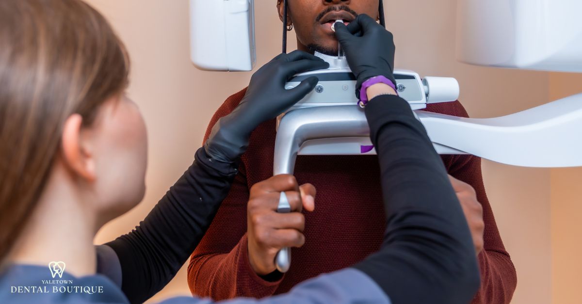
The Scan Itself (20–40 Seconds)
Once you’re positioned, the scan takes less than a minute and requires no contact or discomfort.
- The scanner’s arm rotates once around your head.
- No contact, no pressure, no enclosed “tunnel”, the unit stays fully open.
- You simply stay still while hundreds of images are captured.
3. Review & Treatment Planning
Right after the scan, your dentist reviews the 3D images to guide precise diagnosis and treatment.
- Your dentist reviews the 3D image immediately after the scan.
- Key structures, such as bones, nerves, roots, and sinuses, are assessed in detail.
- The scan becomes the digital roadmap for implants, root canals, extractions, or orthodontic care.
Comfort and Non-Invasiveness
There are no injections or invasive contact involved in a CBCT scan. You may be asked to gently bite on a small positioning stick to help align the laser for accurate imaging.
Beyond that, you simply stay still while the scanner rotates around your head.
Claustrophobia Concerns Addressed
Unlike traditional MRI machines, a CBCT unit is open and non-enclosed. This design helps patients who may feel anxious in tight spaces remain comfortable throughout the scan.
When does a dentist recommend a CBCT scan?
A CBCT scan isn’t used for routine checkups. Your dentist recommends it when 3D clarity meaningfully improves safety, diagnosis, or treatment planning.
This quick guide shows when 3D imaging is essential, optional, or unnecessary, so you know exactly why your dentist may recommend it.
Likely Needed
These situations almost always benefit from 3D imaging:
- Planning a dental implant or bone graft
- Evaluating impacted or wisdom teeth near nerves
- Assessing TMJ joint structure
- Identifying root fractures or complex canal anatomy
- Investigating cysts, lesions, or jaw abnormalities
- Pre-surgical mapping to avoid nerves and sinus cavities
Sometimes Needed
Your dentist may recommend CBCT depending on symptoms, history, and treatment type:
- Persistent unexplained pain with unclear 2D X-rays
- Planning orthodontic treatment or bite correction
- Monitoring previous surgical sites or bone healing
- Assessing sinus involvement for upper molar issues
Not Typically Needed
These situations are usually handled with standard X-rays:
- Routine dental checkups
- Early cavity detection
- Monitoring gum health
- Simple fillings or minor restorations
CBCT vs. Traditional X-Rays: Key Differences
Both CBCT and traditional X-rays play important roles in dentistry, but they are designed for different levels of detail. Understanding their differences explains why your dentist might choose one over the other.
| Feature | Traditional X-Rays | CBCT Scans |
| Image Type | 2D, Flat Images | 3D, Cross-Sectional Views |
| Detail Level | Good for Cavities & Bone Height | High Precision for Bone, Nerves, Sinus |
| Use Cases | Routine Exams, Checkups, Cavities | Implants, Orthodontics, Complex Cases |
| Cost | Lower, Often Fully Covered | Higher, May or May Not Be Covered |
| Radiation | Very Low Exposure | Higher Than X-Ray, Lower Than CT |
2D vs. 3D Imaging
The most fundamental difference is how much dimensional detail each provides:
- Traditional X-Rays: Show flat, two-dimensional images that work well for cavities, bone height, and basic monitoring.
- CBCT: Produces a three-dimensional model of teeth, bone, and soft tissues, giving cross-sectional views from multiple angles.
Diagnostic Detail
Each imaging method offers a different degree of precision:
- Traditional X-Rays: Useful for routine exams but limited in revealing root fractures, nerve pathways, or hidden infections.
- CBCT: Detects complex structures like canal anatomy, sinus boundaries, and bone density, which are crucial for surgical or restorative planning.
Cost and Insurance
Cost is another area where the two differ:
- Traditional X-Rays: Lower in cost and commonly included in standard insurance coverage.
- CBCT: More advanced and therefore more expensive, though many insurance providers offer partial coverage when justified by medical need.
Clinical Use Cases
Knowing when each is appropriate avoids unnecessary imaging:
- Traditional X-Rays: Best for everyday dental care such as checkups, cavity detection, and monitoring periodontal health.
- CBCT: Reserved for complex or high-precision cases, such as implant placement, orthodontic planning, or evaluating unexplained symptoms.

Why We Use CBCT at Yaletown Dental Boutique
For us, CBCT is not about adding another machine to the clinic; it’s about making your treatment safer, clearer, and more predictable. The scan becomes part of how we design care that fits your unique anatomy.
Responsible, Need-Based Use
We follow Health Canada and CDA guidelines, using CBCT only when the added detail directly enhances clinical safety. Fields of view are kept as small as possible and always tailored to the treatment.
Guided, Precision-Based Workflows
CBCT integrates seamlessly into our digital workflow for implants, extractions, TMJ evaluation, orthodontics, and even Invisalign® in Yaletown, helping us map bone structure, root anatomy, and nerve pathways before treatment begins.
Chairside 3D Review for Patients
You see your 3D scan directly at your appointment. We review the anatomy together so you understand what we’re planning and why, reducing uncertainty and improving transparency.
Local Yaletown Convenience
Our in-clinic CBCT system eliminates the need for external imaging centers. Your scan, diagnosis, and treatment planning happen in one place, saving you time and streamlining your care.
Conclusion: Precision That Shapes Better Dentistry
CBCT scans have transformed modern dental care by providing a 3D view of your teeth, jaw, and surrounding structures, helping us accurately diagnose, plan safely, and prevent surprises during treatment. From implants to wisdom tooth surgery, advanced root canals, and TMJ assessments, 3D imaging supports clearer decisions and more predictable outcomes.
If you’re considering a procedure that benefits from precise planning, book a CBCT-guided consultation at Yaletown Dental Boutique.Our team will walk you through your scan, explain your options clearly, and design a treatment plan built on accuracy, safety, and confidence.
Schedule your appointment today and experience the clarity that 3D imaging brings to your care.
Is the scan painful?
No. A CBCT scan is non-invasive and completely painless. You simply sit or stand still while the machine rotates around your head.
How long does it take?
The scan itself lasts less than a minute, with the entire process taking only a few minutes from start to finish.
Is it covered by insurance?
Many dental insurance plans cover CBCT when it’s medically necessary, such as for implant planning or complex diagnosis. Coverage can vary, so checking with your provider is recommended.
Can children have a CBCT scan?
Yes, when clinically appropriate. The scan is safe for children, though dentists only recommend it if the diagnostic benefit clearly outweighs the minimal radiation exposure.
Do I need a CBCT scan for every treatment?
No. CBCT is reserved for cases where extra detail is essential, such as implants, orthodontics, or complex root canals. Routine exams are usually done with traditional X-rays.
How often can a CBCT scan be taken safely?
CBCT is used only when justified. Dentists follow strict ALARA guidelines (“As Low As Reasonably Achievable”) and limit scans to moments when 3D imaging meaningfully improves safety or planning.
Is CBCT more expensive than regular X-rays?
Yes, CBCT is more advanced and may have a higher fee. However, many insurance plans help cover the cost when it’s needed for diagnosis or treatment planning.