At a Glance:
- Dental implant healing happens in stages, beginning with clot formation and inflammation, followed by new blood-vessel growth, bone formation, and ongoing remodelling.
- During the first few weeks, primary mechanical stability gradually gives way to secondary biological stability. Some implants show a temporary stability dip around weeks 3–4, but this pattern is not universal.
- Early woven bone is progressively remodelled into more organized lamellar bone over the following months, while the peri-implant soft tissues form a protective mucosal interface around the implant or abutment.
- Healing time varies according to bone quality, implant stability, surgical conditions, grafting, loading protocol, and individual biology. Crown placement is based on clinical readiness rather than a fixed calendar date.
- Increasing pain, worsening swelling, drainage, persistent bleeding, or suspected implant mobility should be assessed professionally rather than monitored through self-testing.
Dental implant healing involves much more than waiting for soreness and swelling to disappear. Beneath the gum, the surgical site moves through overlapping stages of blood clot formation, inflammation, new blood-vessel development, bone formation, and ongoing remodelling around the implant surface.
This dental implant healing timeline is important because the implant does not become biologically integrated with the jawbone immediately after placement. Its initial stability comes largely from mechanical engagement with the existing bone.
Over the following weeks and months, healing tissues gradually create biological stability as new bone forms and remodels at the implant-bone interface.
The process is not identical for every patient or implant site. Bone quality, surgical conditions, implant design, healing capacity, grafting procedures, and loading can all influence the rate and pattern of healing.
What feels quiet from the outside can therefore represent an active period of cellular and structural change beneath the surface.
The Immediate Cellular Response: Clot Formation, Inflammation, and Early Angiogenesis (Days 1–3)
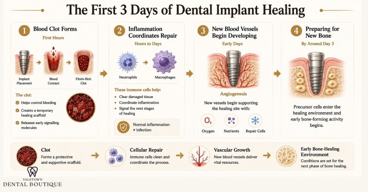
The first few days after dental implant placement are often associated with swelling, tenderness, and minor bleeding. At the microscopic level, however, the surgical site is already undergoing a coordinated sequence of events that prepares the area for new bone formation.
These processes overlap rather than occurring as separate steps on a fixed schedule.
First Hours: Blood Contact and Clot Formation
Implant placement creates a controlled bone wound. As soon as the implant enters the prepared site, its surface comes into contact with blood and tissue fluid.
Proteins rapidly adsorb to the implant surface, platelets become activated, and the coagulation system forms a fibrin-rich blood clot within the spaces between the implant and surrounding bone.
This clot does more than control bleeding. Its fibrin network acts as a temporary biological scaffold through which inflammatory, vascular, and bone-forming precursor cells can later migrate.
Platelets within the clot also release signalling molecules and growth factors that participate in the early healing response.
The Early Inflammatory Response
Inflammation is a normal component of surgical wound healing and does not, by itself, mean that an implant site is infected.
During the first days, immune cells enter the area and help clear damaged tissue and coordinate the next stages of repair.
Neutrophils are prominent early in this response, followed by increasing macrophage activity.
Macrophages have several roles. In addition to clearing cellular debris, they participate in signalling pathways that influence inflammation, blood-vessel formation, and the recruitment and differentiation of cells involved in bone repair.
The inflammatory response therefore needs to be regulated rather than simply eliminated. It is part of the transition from the initial surgical wound toward tissue regeneration.
Angiogenesis Creates a Blood Supply for Repair
New bone cannot form without an adequate blood supply.
During early healing, signalling molecules such as vascular endothelial growth factor (VEGF) contribute to angiogenesis, the process through which new blood vessels develop from existing vessels.
These vessels provide oxygen, nutrients, and access for cells involved in tissue repair. Vascular development also occurs alongside the migration of mesenchymal and osteogenic precursor cells into the healing site.
This close relationship between vascular growth and bone formation is one reason the earliest stages of implant healing are biologically active even before meaningful new bone has formed around the implant.
Preparing the Implant Surface for New Bone
As early healing progresses, precursor cells can migrate through the provisional fibrin matrix and differentiate toward bone-forming cell lineages.
Osteoblasts are the cells responsible for producing new bone matrix. Their activity eventually contributes to new bone developing both from the surrounding bone toward the implant and, depending on implant surface characteristics and local conditions, at or near the implant interface.
This is the beginning of the transition from a blood-clot-filled surgical site to a biologically active bone-healing environment.
Importantly, this does not mean the implant is already osseointegrated during the first few days. Its stability at this stage still depends predominantly on the mechanical engagement established at surgery.
What is happening by the end of day 3?
By approximately the first several days, the surgical site is moving from haemostasis and acute inflammation toward the proliferative stages of wound healing.
At this point:
- The initial fibrin clot has provided a temporary healing matrix
- Inflammatory cells are coordinating debris removal and repair
- New vascular development is beginning
- Osteogenic precursor cells are being recruited to the healing environment
- Early signals for new bone formation are becoming active
These events form the biological foundation for the next phase of the dental implant healing timeline, when the relationship between mechanical implant stability and newly developing biological stability begins to change.

The Biomechanical Pivot: Understanding the Stability Dip (Weeks 2–4)
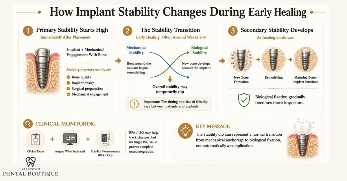
One of the less visible stages of the dental implant healing timeline occurs during the first several weeks after placement. The implant can feel completely uneventful from the patient’s perspective while the way it is anchored within the jaw is changing beneath the surface.
Immediately after placement, stability comes predominantly from mechanical engagement between the implant and the surrounding bone. As healing progresses, this primary stability gradually gives way to secondary stability, which develops through new bone formation and remodelling at the implant surface.
The transition can be associated with a temporary reduction in measured implant stability, commonly referred to as the dental implant stability dip.
Primary vs. Secondary Implant Stability
Although both contribute to a stable implant, primary and secondary stability arise through different mechanisms.
Primary implant stability is mechanical. It is established at surgery through engagement between the implant and the prepared bone site.
The amount of primary stability can be influenced by factors such as:
- Local bone density and architecture
- Implant geometry and dimensions
- Surgical site preparation
- Implant position
- The degree of mechanical engagement achieved during placement
Secondary implant stability develops biologically. As healing proceeds, newly formed bone develops at and around the implant interface and subsequently remodels and matures.
Primary stability therefore describes the implant’s initial mechanical anchorage, while secondary stability reflects the developing biological fixation associated with osseointegration.
Why can implant stability temporarily decrease?
The bone immediately surrounding an implant does not remain unchanged after surgery.
Some of the bone involved in the implant’s initial mechanical engagement undergoes remodelling as part of the normal healing response. At the same time, newly formed bone is developing but may not yet have matured sufficiently to provide the biological stability seen later in healing.
This creates an important transition:
Mechanical stability ↓
Biological stability ↑
If the reduction in initial mechanical support temporarily occurs faster than secondary biological stability develops, measurements of overall implant stability may decrease before increasing again.
That transitional pattern is what is commonly described as the stability dip.
Does the stability dip always occur between weeks 2 and 4?
A stability dip can occur during early healing, with implant stability often decreasing around three to four weeks after placement before increasing again as biological stability develops.
However, this pattern is not universal. Some implants show a noticeable reduction in measured stability, while others demonstrate a smaller decline, an earlier or later change, or relatively stable measurements throughout the healing period.
For that reason, the Weeks 2–4 timeframe is best treated as a commonly observed transition period rather than a fixed biological rule. Stating that every implant reaches its lowest total stability at week 3 or 4 would overstate the available evidence.
The pattern can vary according to factors such as:
- Bone quality and implant location
- Initial implant stability
- Implant macrogeometry and surface characteristics
- Surgical technique
- Healing conditions
- Loading protocol
- Patient-specific biological factors
Why Mechanical Loading Matters During Early Healing
Bone formation around an implant occurs in a mechanical environment. Some mechanical stimulation can be compatible with osseointegration, and selected implants can even be restored using immediate or early loading protocols when clinical conditions are appropriate. The concern is excessive movement at the bone-implant interface during healing.
Experimental evidence indicates that excessive interfacial micromotion can interfere with bone formation and favour a fibrous tissue interface rather than stable osseointegration.
Research has not established one universal micromotion threshold that applies to every implant and clinical situation.
Patients therefore should not intentionally test an implant by pushing it with the tongue or fingers, and chewing or loading instructions should follow the treatment plan provided for the specific implant.
An implant restored immediately may have different loading instructions from one intentionally left unloaded during healing.
How do dentists assess implant stability?
Implant stability cannot be determined simply by how the area feels to the patient.
Depending on the clinical situation, dentists may assess healing through examination, the restorative timeline, radiographic findings, and mechanical stability measurements.
One non-invasive method is resonance frequency analysis (RFA). The measurement is commonly expressed as an Implant Stability Quotient (ISQ).
ISQ values can help clinicians track changes in mechanical implant stability over time. However, an ISQ number is not a direct measurement of microscopic bone-to-implant contact, and there is no single value that independently proves that an implant is fully osseointegrated or ready for restoration.
Insertion torque and ISQ also measure different aspects of implant stability and should not be treated as interchangeable measurements.
The stability dip is a transition, not automatically a complication
A temporary decline in measured stability during early healing can reflect the transition from mechanical anchorage to biological fixation rather than implant failure.
The Weeks 2–4 period is therefore useful as a clinical reference point, but it should not be interpreted as a universal window of instability. Dentists evaluate implant healing in the context of the surgical procedure, bone conditions, symptoms, clinical findings, imaging when indicated, and the planned loading protocol.
The important biological milestone is not reaching a particular calendar week. It is the progressive development of a stable bone-implant interface capable of supporting the next phase of treatment.
The Architecture of Osseointegration: Woven Bone to Lamellar Bone (Months 1–3)
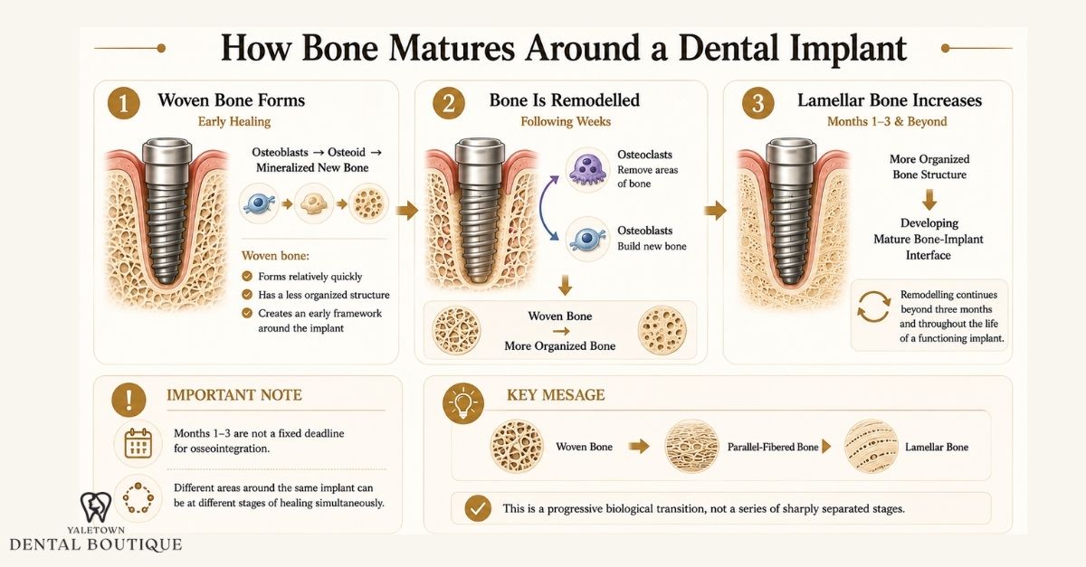
After the earliest inflammatory and stability-transition phases, healing around a dental implant increasingly shifts toward new bone formation and remodelling.
This process is called osseointegration, generally referring to a direct structural and functional connection between living bone and the implant surface. It does not occur all at once. The bone immediately surrounding an implant changes in composition and organization as healing progresses.
One of the most important transitions is the progression from rapidly formed woven bone toward more organized parallel-fibered and lamellar bone.
Phase 1: Woven bone creates the early framework
New bone formation can begin surprisingly early. Histologic studies have identified newly formed bone at or near implant surfaces within the first weeks of healing, although the timing and amount vary according to the implant surface, bone characteristics, surgical site, and other factors.
The earliest newly formed bone is predominantly woven bone.
Woven bone is produced relatively quickly and has a less organized collagen architecture than mature lamellar bone. Its rapid formation makes it useful during early repair because it can begin filling spaces between the existing bone and implant surface while the healing site is still developing.
Osteoblasts produce an initially unmineralized organic matrix called osteoid. This matrix subsequently mineralizes, contributing to newly formed bone around the implant.
Bone can develop from the existing bone surfaces toward the implant, and new bone formation may also occur at or near appropriately prepared implant surfaces. Together, these processes progressively increase the amount of bone associated with the implant interface.
Phase 2: Woven bone is remodelled into more organized bone
Woven bone is an early repair tissue, not the final architecture surrounding a functioning implant.
Over the following weeks and months, bone remodelling progressively replaces portions of this immature structure with more organized parallel-fibered and lamellar bone.
This remodelling involves coordinated activity between:
- Osteoclasts, which resorb areas of existing bone
- Osteoblasts, which produce new bone matrix
- Osteocytes and other signalling cells involved in regulating bone adaptation and turnover
Lamellar bone has a more organized structural arrangement than woven bone and contributes to the mature architecture that supports the implant over time.
Importantly, this is not a simple one-way event in which all woven bone disappears at a specific date. Different regions around an implant may be at different stages of formation and remodelling at the same time.
When does lamellar bone begin to form?
The Months 1–3 period captures an important portion of this transition, but it should not be interpreted as a universal deadline for osseointegration.
Experimental and human histologic studies show that early bone formation can begin within the first week and becomes increasingly evident during the following several weeks. Reviews of implant healing describe substantial bone remodelling beginning approximately 6 to 12 weeks after placement, although the exact timing varies.
By this stage, increasing amounts of more organized bone may be present around the implant. However, bone maturation and remodelling continue after three months and can continue throughout the life of a functioning implant.
For this reason:
Woven bone → parallel-fibered and lamellar bone
is better viewed as a progressive biological transition than as two sharply separated stages.
Does chewing turn woven bone into lamellar bone?
Mechanical loading influences how bone adapts and remodels, but it would be inaccurate to say that normal chewing is what directly converts woven bone into lamellar bone.
Bone maturation is governed by the broader biological remodelling process. The local mechanical environment can influence that process, which is one reason loading protocols are selected according to the circumstances of the individual implant.
Some implants may be restored or loaded relatively early when appropriate stability and clinical conditions are present. Others are intentionally given a longer healing period before substantial functional loading.
There is therefore no universal rule that an implant must remain completely unloaded for three months before osseointegration can occur.
Can X-rays confirm osseointegration?
Dentists may use clinical examination together with radiographic imaging assessments to evaluate the tissues around an implant and look for findings such as changes in peri-implant bone levels or other abnormalities.
However, a conventional dental radiograph cannot directly show the microscopic percentage of bone-to-implant contact.
Similarly, a normal-looking X-ray does not by itself prove that complete histologic osseointegration has occurred. Clinical decisions about loading or restoration can incorporate several findings, including implant stability, symptoms, examination findings, healing time, surgical conditions, and imaging when indicated.
This distinction is important because osseointegration is a microscopic biological process, while the tools available during routine clinical care assess it largely through indirect indicators.
What happens after three months?
Three months is not the biological endpoint of implant healing.
By this stage, many implants may have developed substantial secondary stability and more mature bone at the implant interface, but the amount and maturity of that bone vary between cases.
Bone surrounding a functioning implant remains dynamic. It continues to undergo remodelling in response to normal physiological turnover and its mechanical environment long after the initial healing period has ended.
The transition from woven bone to increasingly organized lamellar bone therefore represents the development of a more mature implant-bone interface, not the completion of all biological activity around the implant.
The Soft Tissue Barrier: Formation of the Peri-Implant Mucosal Seal
Osseointegration focuses on the relationship between the implant and bone, but successful healing also depends on the soft tissue that develops around the portion of the implant or abutment extending through the gums.
This tissue is called the peri-implant mucosa. Once established, it forms a protective biological interface between the oral environment and the deeper tissues surrounding the implant.
That barrier is important because the implant passes through an environment containing saliva, food debris, and a complex oral microbiome. The peri-implant mucosa helps separate these external influences from the underlying connective tissue and supporting bone.
How does the gum attach around an implant?
The soft tissue surrounding an implant includes an epithelial component and an underlying connective-tissue component.
Near the implant or abutment surface, epithelial cells form a peri-implant junctional or barrier epithelium. These cells can adhere to the implant surface through an internal basal lamina and structures known as hemidesmosomes.
Hemidesmosomes act as microscopic anchoring structures between epithelial cells and the underlying surface.
This creates an epithelial attachment that shares some features with the junctional epithelium around a natural tooth. However, the comparison has important limits.
An implant does not have the same soft tissue attachment as a natural tooth
Around a natural tooth, connective-tissue collagen fibres can insert into the root cementum, forming part of the periodontal attachment apparatus.
A dental implant has no cementum and no periodontal ligament. Instead, collagen fibres within the peri-implant connective tissue tend to run parallel or circumferentially to the implant surface rather than inserting directly into it in the same way as fibres around a natural tooth.
The peri-implant connective tissue also tends to contain:
- A higher proportion of collagen
- Fewer fibroblasts in some regions
- A different vascular supply
- No blood vessels originating from a periodontal ligament
These anatomical differences mean that the peri-implant mucosal seal should not be described as an exact reproduction of the natural periodontal attachment.
Why Blood Supply Matters
Natural periodontal tissues receive vascular contributions from several sources, including vessels associated with the periodontal ligament. Because the implant fixture itself has no periodontal ligament, the peri-implant soft tissues rely more heavily on blood vessels from the surrounding supraperiosteal tissues.
Adequate vascularity is important during healing because oxygen, nutrients, immune cells, and signalling molecules all depend on the local circulation.
The vascular network therefore contributes both to initial wound healing and to the long-term maintenance of the peri-implant tissues.
The mucosal seal is a biological defence, not an impermeable barrier
It can be tempting to describe healthy peri-implant tissue as creating a completely sealed barrier against bacteria. Biologically, that would be too strong.
The peri-implant mucosa forms an important protective interface, but it is not impermeable.
Plaque biofilm can accumulate around implant-supported restorations just as it can around natural teeth. If plaque-induced inflammation develops in the soft tissue surrounding an implant, the condition is called peri-implant mucositis. Clinical signs can include bleeding on gentle probing, redness, or swelling.
Peri-implantitis is a different condition in which inflammation around an implant is accompanied by progressive loss of supporting bone beyond the changes expected from initial bone remodelling.
Maintaining access for effective plaque removal therefore remains important after the implant and its restoration have healed.
Why the Restoration Above the Implant Matters
The peri-implant mucosa does not develop independently of the components passing through it.
The shape and position of the healing abutment and, later, the definitive restoration can influence the contour of the surrounding soft tissue and how easily the area can be cleaned.
The transition from the implant platform toward the crown is often described as the emergence profile.
A restoration must therefore do more than replace the visible tooth. Its contours also need to be considered in relation to the surrounding mucosa, access for oral hygiene, and the overall restorative plan.
Soft tissue healing continues alongside bone healing
Soft tissue closure generally occurs much sooner than the deeper process of osseointegration, but the appearance of a healed gum surface does not mean that all biological maturation beneath it has finished.
As the peri-implant mucosa matures, its epithelial and connective-tissue components establish the interface that helps protect the deeper implant-bone environment.
Long-term maintenance then becomes important. The Canadian Dental Association advises that implants, like natural teeth, need regular cleaning and dental checkups so the surrounding tissues and restoration can be monitored.
The peri-implant mucosal seal is therefore best viewed as a living biological interface that requires ongoing maintenance, rather than as a permanent barrier that becomes complete once the gum has closed.
Expected Milestones: The Biological Progress Map
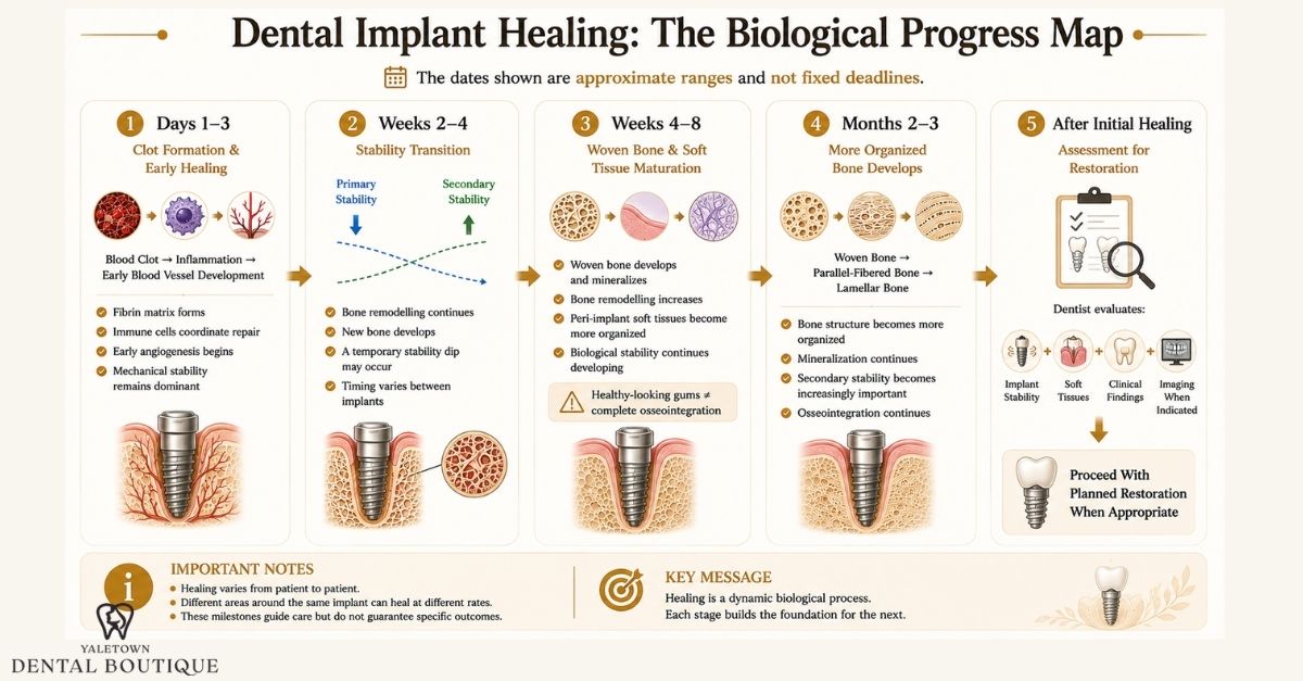
Dental implant healing does not follow a perfectly fixed schedule, but several biological milestones tend to occur in a recognizable sequence. The timeline below shows how the surgical site may progress from early clot formation and inflammation to increasing bone organization, soft tissue maturation, and eventual assessment for the restorative phase.
The exact timing can vary according to the implant site, bone quality, surgical technique, grafting, implant stability, loading protocol, and individual healing response.
Days 1–3: Hemostasis and Fibrin Networking
Main biological activity: Blood clot formation, inflammation, and early vascular signalling
Immediately after implant placement, blood fills microscopic spaces within the surgical site and contacts the implant surface. Platelets become activated and a fibrin-rich clot forms, creating a provisional matrix for the early healing response.
During the following days:
- Neutrophils and macrophages participate in the inflammatory response
- Damaged tissue and cellular debris begin to be cleared
- Signalling molecules coordinate tissue repair
- Early angiogenesis begins to establish vascular pathways
- Osteogenic precursor cells begin participating in the developing healing environment
The implant is not biologically integrated at this stage. Its stability still depends predominantly on the mechanical engagement established during surgery.
Weeks 2–4: The Biomechanical Stability Pivot
Main biological activity: Declining contribution from primary stability while secondary biological stability develops
During early bone remodelling, some of the bone contributing to the implant’s initial mechanical anchorage is remodelled. At the same time, newly forming bone is beginning to contribute to biological fixation.
This changing relationship can produce a temporary implant stability dip.
Some clinical studies have reported lower stability measurements at approximately three to four weeks, but this pattern is not universal. Other implants show smaller changes, different timing, or no distinct stability dip.
During this period:
- Primary mechanical stability may decrease
- New bone formation continues around the implant
- Secondary biological stability begins contributing more substantially
- Early peri-implant connective tissue and epithelial organization continues
- Excessive movement at the bone-implant interface remains undesirable
Weeks 2–4 should therefore be viewed as a commonly observed transition period, not a universal point when every implant is at its weakest.
Weeks 4–8: Woven Bone Consolidation and Soft Tissue Maturation
Main biological activity: Increasing new bone formation and development of the peri-implant mucosal interface
By this phase, immature woven bone can occupy increasing portions of the healing interface.
Woven bone forms relatively quickly and provides an early framework that is subsequently remodelled into more organized bone.
At the same time, the soft tissues surrounding the transmucosal portion of the implant continue to mature.
Preclinical histologic studies indicate that peri-implant epithelial and connective-tissue attachment develops progressively over several weeks, with a more mature soft-tissue interface observed at approximately 6–8 weeks in experimental models. This timeframe should not be interpreted as a fixed clinical healing timeline for individual patients.
During this period:
Woven bone continues to develop and mineralize
- Bone remodelling becomes increasingly active
- Secondary implant stability may continue to increase
- The peri-implant epithelial barrier becomes more organized
- Connective-tissue collagen develops around the implant or abutment
- Surface gum healing may appear advanced even though deeper bone maturation continues
The appearance of healthy gum tissue does not mean that osseointegration is complete.
Months 2–3: Increasing Bone Organization and Secondary Stability
Main biological activity: Remodelling of early bone toward more organized parallel-fibered and lamellar bone
As healing continues, portions of the rapidly formed woven bone are remodelled into more structurally organized bone.
Osteoclasts remove selected areas of existing or newly formed bone while osteoblasts deposit new bone matrix. Through repeated remodelling, the architecture around the implant becomes progressively more mature.
During this phase:
Woven bone is progressively remodelled
- Parallel-fibered and lamellar bone become increasingly represented
- Mineralization and structural organization continue
- Biological fixation contributes increasingly to implant stability
- Clinical and radiographic findings may be reviewed before changing the loading or restorative plan
This is an important period of osseointegration, but three months is not a universal biological endpoint.
After the Initial Healing Period: Assessment for Restoration
Main clinical question: Is this implant ready for the planned restorative phase?
There is no single calendar date when every implant becomes ready for a permanent crown.
The timing can depend on:
- Primary and secondary implant stability
- Implant location
- Bone quality and quantity
- Whether bone grafting was required
- Surgical and implant characteristics
- Soft-tissue condition
- Symptoms and clinical findings
- Loading protocol
- The type of restoration being planned
Modern implant dentistry includes immediate, early, and conventional loading protocols in appropriately selected cases. This means that placing a restoration does not always require waiting three to six months, while other clinical situations may require a longer healing interval.
When appropriate, dentists may use clinical examination, stability assessment, and radiographic findings to help determine how healing is progressing. Imaging can provide information about peri-implant bone, but it cannot directly demonstrate microscopic bone-to-implant contact.
Once the implant and surrounding tissues are considered suitable for the planned restorative phase, treatment can proceed toward the definitive dental implant restoration.
Even after a crown is placed, bone surrounding a functioning implant remains biologically active and continues to remodel over time.
Why Dental Implant Healing Times Vary
The biological sequence of implant healing is broadly similar between patients, but its timing and clinical expression can vary substantially. Osseointegration is influenced by both the local environment around the implant and the patient’s overall capacity for wound healing and bone remodelling.
Bone Quality and Local Anatomy
Bone density and architecture affect the mechanical stability achieved when an implant is placed and can influence the subsequent healing environment. Implant literature has traditionally described bone using classifications ranging from dense cortical bone to lower-density trabecular bone, sometimes referred to as Types I through IV. However, these categories are descriptive rather than exact biological measurements, and different classification methods do not always produce directly comparable results.
Lower-density bone can provide less initial mechanical engagement in some situations, while denser bone presents different surgical and vascular considerations. Implant geometry, site preparation, anatomical location, and the amount and distribution of cortical and trabecular bone therefore need to be considered together rather than relying on a bone-type label alone.
Blood Supply and Healing Environment
Bone formation requires vascular support. Blood vessels deliver oxygen, nutrients, immune cells, and signalling molecules to the healing site, so local vascularity is closely linked with tissue repair and new bone formation. Surgical trauma, grafting, local anatomy, and other factors can alter the environment in which angiogenesis and bone regeneration occur.
Systemic and Patient-Specific Factors
General health can also influence healing, although having a systemic condition does not automatically prevent successful implant treatment. For example, evidence indicates that glycaemic control is clinically relevant in patients with diabetes, while smoking can adversely affect vascularity, wound healing, and implant outcomes. The effects of other diseases and medications are more variable and should be considered individually rather than generalized across all patients.
Surgical and Loading Factors
Healing can also differ according to whether grafting was performed, the stability achieved at placement, implant design and surface characteristics, and whether the implant follows an immediate, early, or conventional loading protocol. For this reason, the appropriate restorative timeline is determined from the overall clinical picture rather than from the number of weeks that have passed since surgery.
Clinical Red Flags: Normal Surgical Discomfort vs. Structural Implant Failure
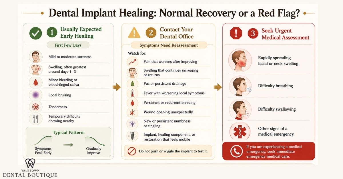
Some soreness, swelling, and minor bleeding are expected after dental implant surgery. What matters most is the direction of recovery. In an uncomplicated course, symptoms generally become less noticeable as the first several days pass.
Persistent or worsening symptoms deserve closer attention because they can indicate infection, wound-healing problems, mechanical complications, or difficulty establishing osseointegration.
What Usually Falls Within Normal Early Healing?
Postoperative experiences vary with the number and location of implants, the extent of surgery, grafting, flap design, and individual healing response.
After uncomplicated implant placement, it can be normal to experience:
- Mild to moderate soreness during the first few days
- Swelling that becomes most noticeable during approximately the first 1–3 days
- Minor bleeding or blood-tinged saliva early after surgery
- Local bruising
- Tenderness around the surgical site
- Temporary difficulty chewing near the treated area
Pain is most significant during the early postoperative period and then declines. Swelling often reaches its greatest level around the second or third day before gradually decreasing.
The exact pattern is not identical for every patient, particularly when implant placement is combined with bone grafting or more extensive surgery.
When Pain Deserves Reassessment
Pain alone does not establish that an implant has failed.
The trend and severity provide more useful information. Pain that is mild or gradually improving during the first several days is generally more consistent with routine postoperative healing.
Contact the treating dentist if pain:
- Becomes more intense after initially improving
- Remains moderate to severe beyond the early postoperative period
- Develops alongside increasing swelling
- Is associated with pus, fever, an unpleasant taste, or wound drainage
- Is substantially different from the postoperative course your dentist described
One observational study found an association between moderate-to-severe pain persisting beyond 72 hours and subsequent failure of osseointegration. That finding does not mean persistent pain proves implant failure. Pain has many possible causes, and the study design cannot establish a universal diagnostic rule.
When Swelling May Be More Concerning
Swelling is a normal inflammatory response to surgery and commonly becomes more noticeable during the first few days. The more important question is what happens afterward.
Professional assessment may be appropriate when swelling:
- Continues increasing rather than beginning to settle
- Persists substantially longer than expected for the procedure
- Returns after having improved
- Is accompanied by increasing pain, fever, pus, or spreading redness
- Makes it increasingly difficult to open the mouth
Rapidly spreading facial or neck swelling, particularly when accompanied by difficulty breathing or swallowing, requires urgent medical assessment rather than routine dental follow-up.
What If the Implant Feels Loose?
A successfully integrated dental implant should not demonstrate detectable clinical mobility.
However, patients should not push, wiggle, or apply horizontal pressure to an implant to test its stability. Deliberately loading a healing implant provides no reliable home assessment and may disturb the surgical site.
A sensation of movement also does not automatically mean that the implant fixture itself has moved. In a restored or immediately provisionalized implant, movement can sometimes come from another component, such as a healing abutment, screw, temporary crown, or definitive restoration.
If any part of the implant restoration appears or feels mobile, the appropriate response is to have it examined clinically.
True implant mobility can be an important sign of failed or lost osseointegration, but that diagnosis is made by the dentist rather than through patient self-testing.
Other Signs That Should Prompt a Dental Review
Contact the treating dental office if you develop:
- Pus or persistent drainage from the surgical site
- Increasing redness or swelling after the initial recovery period
- Persistent or recurrent bleeding that does not respond to the postoperative instructions provided
- A wound that opens significantly or exposes material unexpectedly
- Persistent bad taste or malodour associated with other signs of infection
- New or persistent numbness, tingling, or altered sensation
- Movement of the implant, healing component, or restoration
- Pain or swelling that begins worsening after a period of improvement
Possible complications associated with dental implant treatment can include bleeding, infection, altered sensation or numbness, and failure of the implant to successfully integrate with the surrounding bone.
How is a suspected implant complication evaluated?
A dentist does not diagnose early implant failure from pain alone.
Assessment may include:
Symptom history: When the problem began, whether it is improving or worsening, and whether there is pain, swelling, drainage, bleeding, or altered sensation.
Clinical examination: The surgical site, surrounding soft tissue, healing components, occlusion, and implant or prosthetic components can be evaluated.
Stability assessment when appropriate: The clinician may assess for detectable mobility or use other implant-stability measurements depending on the stage of treatment.
Radiographic assessment when indicated: Dental imaging may be used to investigate peri-implant bone, implant position, radiolucency, or other suspected problems. Imaging complements the clinical examination but does not independently diagnose microscopic osseointegration.
Depending on the findings, management can range from observation and modification of postoperative care to treatment of an infection or mechanical complication. Failure of osseointegration may require a different approach, including implant removal in some circumstances.
The Clinical Baseline for Triage
During early healing, soreness and swelling are generally most noticeable in the first few days and should gradually improve. Professional reassessment is appropriate if pain or swelling begins increasing instead of settling, drainage or other signs of infection develop, bleeding remains difficult to control, or the implant or an associated component appears mobile.
Patients should not try to test implant stability by pushing or wiggling the area, since a dentist needs to determine which component is moving and why.
Symptoms suggesting a significant dental complication may warrant immediate clinical triage. Rapidly spreading swelling, difficulty breathing or swallowing, or another medical emergency requires urgent medical assessment.
Conclusion
Dental implant healing happens in overlapping stages, from early clot formation and inflammation to the shift from primary to secondary stability, osseointegration, and ongoing bone remodelling.
Although discomfort often settles within days, deeper healing continues for weeks or months. At Yaletown Dental Boutique, readiness for the next stage is assessed based on factors such as implant stability, soft-tissue health, clinical findings, and imaging when indicated.
Once healing is appropriate, treatment can progress toward the final dental implant restoration, such as a crown. The timeline is best viewed as a biological progression rather than a fixed countdown to a specific week or month.
Is swelling normal after dental implant surgery?
Yes. Swelling is common and often becomes most noticeable during the first two to three days before gradually improving. Contact your dentist if swelling keeps worsening or occurs with fever, increasing pain, or drainage.
How long does pain last after dental implant surgery?
Discomfort is usually greatest during the first 24–72 hours and should gradually improve. Pain that intensifies, persists significantly, or occurs with swelling or drainage should be assessed.
When can I eat normally after a dental implant?
There is no fixed timeline. Softer foods are usually recommended early on, with firmer foods added as comfort improves and according to your dentist’s instructions.
When can I return to work after implant surgery?
Some patients return quickly, while others need more recovery time. It depends on the extent of surgery, discomfort, medications, and the physical demands of your job.
How long does osseointegration take?
Osseointegration develops gradually over weeks and months. The timeline varies with the implant site, bone conditions, stability, grafting, and the loading protocol used.
What is the dental implant stability dip?
The stability dip is a temporary decrease in measured implant stability that can occur as mechanical stability declines and biological stability develops. It is often reported around weeks three to four, but this timing is not universal.
How do I know if my dental implant has integrated with the bone?
You cannot reliably judge osseointegration by how the implant feels. Dentists assess healing using clinical findings, implant stability when appropriate, healing time, and imaging when indicated.
What does it mean if my dental implant feels loose?
A loose sensation should be assessed, but it does not always mean the implant itself is moving. An abutment, screw, or restoration can also become loose. Do not test the implant by wiggling it.
When is the permanent crown placed after an implant?
Timing varies. The dentist considers implant stability, bone and soft-tissue healing, grafting, implant location, and the selected loading protocol before placing the final restoration.
What are signs that implant healing may need professional assessment?
Contact your dentist if pain or swelling is worsening, bleeding remains difficult to control, pus or drainage develops, or the implant or restoration feels mobile. Rapid swelling with breathing or swallowing difficulty requires urgent medical care.